Spinal Endoscope & Minimally Invasive Surgical Instruments Manufacturer | QuNaMai
Wishlist

{{ variable.name }}


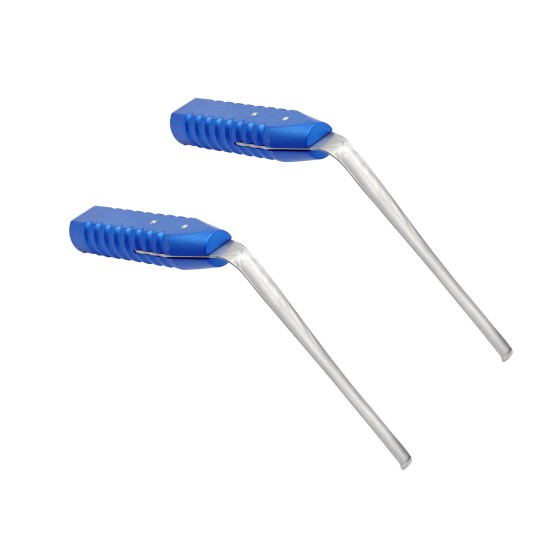
Structural features and advantages
Size advantage
The 2MM diameter makes the nerve retractor have excellent passability during UBE surgery. In spinal surgery, the surgical passage is relatively narrow, especially in the cervical and thoracic regions. The smaller size enables the nerve retractor to smoothly enter the surgical site without causing excessive compression on the surrounding tissues. This helps to approach the nerve structures within a limited space, such as flexibly extending around the nerves when operating on vulnerable areas like the cervical foramen or thoracic canal.
Advantages of ball head design
Reduce the risk of nerve injury: The ball head design is a key feature of the nerve retractor. The surface of the ball head is smooth without sharp edges, which can minimize the risk of nerve cutting or contusion when separating and pulling nerve tissue. In spinal surgery, nerve tissues are very fragile, such as the spinal cord and nerve roots. Such a design can safely separate the nerves from the surrounding tissues and avoid iatrogenic nerve damage caused by instruments.
Good tissue adaptability: The ball head can better fit the irregular shapes of the nerve surface and surrounding tissues. When dealing with adherent tissues around the nerve or searching for nerve pathways in complex anatomical structures, the ball head can operate along the nerve contour, improving the accuracy and efficiency of separation. For instance, in the surgery for lumbar intervertebral disc protrusion, the bulb of the nerve retractor can be gently separated from the protruding intervertebral disc tissue along the direction of the nerve root.
Application scenarios in UBE surgery
Application in the surgery of intervertebral disc protrusion
In UBE surgery for lumbar intervertebral disc protrusion or cervical intervertebral disc protrusion, a nerve retractor is used to protect and retract the nerve roots. After locating the protruding nucleus pulposus and the compressed nerve root under an endoscope, a 2MM bulbar nerve retractor is gently inserted between the nerve and the nucleus pulposus from the side or bottom. The bulbous head can fit the shape of the nerve root and be slowly pulled apart, allowing the doctor to see the protruding nucleus pulposus more clearly while avoiding damage to the nerve root. For instance, in common intervertebral disc protrusion segments such as L4-L5 or C5-C6, the nerve retractor not only ensures the safety of the nerve roots but also creates favorable operating conditions for nucleus pulposus removal surgery, effectively alleviating the nerve compression symptoms in the lower or upper limbs of patients.
The application of spinal canal decompression surgery
In UBE surgery for spinal stenosis, a nerve retractor can be used to separate the nerve tissue within the spinal canal from the surrounding compressive tissues, such as the ligamentum flavum and osteophytes. During cervical or lumbar spinal canal decompression surgery, when dealing with the ligamentum flavum that has proliferated on the posterior wall of the spinal canal, a nerve retractor can gently pull apart the spinal cord or nerve roots located beneath the ligamentum flavum to prevent nerve damage during the removal of the ligamentum flavum. For lateral recess stenosis caused by osteophyte formation on the lateral wall of the spinal canal, a nerve retractor also helps to separate the nerve from the bone, providing safety for subsequent bone removal surgery.
The application of spinal trauma surgery
In trauma surgeries such as spinal fractures and dislocations, nerve retractors can be used to examine and protect nerve tissues. For instance, in surgeries involving thoracic vertebrae fractures and those with a risk of spinal cord injury, a nerve retractor can carefully examine the condition around the spinal cord before fracture reduction and fixation. It can separate bone fragments or hematoma tissues that may compress the spinal cord from the spinal cord and gently pull the spinal cord apart with the retractor to ensure the safety of the spinal cord during fracture reduction.
Operation precautions and skills
Precautions for Operation
Visual operation is of vital importance: UBE surgery relies on the visibility of the endoscope. Before using a nerve retractor, it is essential to ensure a clear endoscopic field of view and accurately determine the relationship between the retractor and surrounding nerves, blood vessels and other tissues. Any blind operation may lead to serious consequences such as nerve damage.
Gentle operation principle: Due to the fragility of nerve tissue, the principle of gentleness must be followed during operation. When inserting and pulling out the nerve retractor, excessive force should be avoided to prevent nerve damage from pulling. Especially when the nerve is already under compression or damage, the operation needs to be carried out with even greater caution.
Pay attention to the position of the nerve retractor: During the operation, be sure to keep the position of the nerve retractor stable to avoid accidental movement during the surgical operation (such as tissue resection, irrigation, etc.) that may damage the nerve. If the position of the nerve retractor needs to be adjusted, it should be done slowly under direct vision.
Operation skills
Insertion technique: Based on the anatomical path and nerve position observed by the endoscope, slowly insert the nerve retractor at an appropriate Angle and direction. During the insertion process, take advantage of its 2MM diameter and carefully enter the area around the nerve along the natural anatomical gap or the established working channel to avoid forced insertion that may damage the surrounding tissues. For instance, during anterior cervical spine surgery, when inserting a nerve retractor, it can first be inserted along the gap at the anterior edge of the vertebral body, and then the direction should be slowly adjusted to approach the nerve root.
Traction technique: When pulling the nerve, apply force gently and continuously. First, make sure the ball head is in full contact with the nerve surface, and then gradually apply tension to gradually separate the nerve from the surrounding tissues. The direction of traction should be determined based on the anatomical direction of the nerve and the requirements of the surgical operation. Generally, the nerve should be pulled along the natural extension direction to avoid twisting or excessive bending of the nerve. At the same time, combined with real-time endoscopic observation, the traction intensity and Angle should be flexibly adjusted according to neural responses and changes in surrounding tissues.
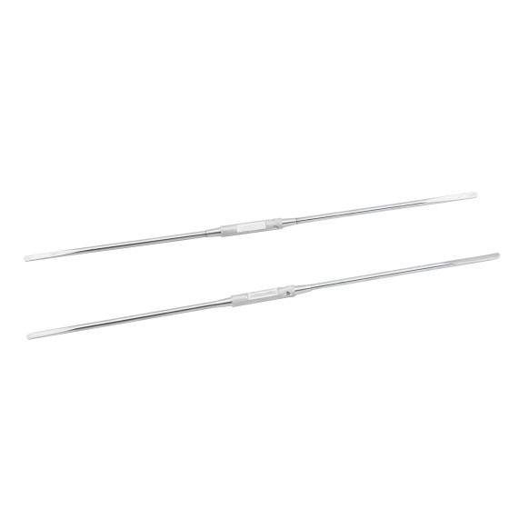
| Surgical instruments | |
| Material | Stainless steel |
| Cryogenic plasma | Autoclaving |

{{ item.content }}

{{ item.created_at_format }}
{{ item.reply }}
To find out more about our products and solutions, please fill out the form below and one of our experts will get back to you shortly.