Spinal Endoscope & Minimally Invasive Surgical Instruments Manufacturer | QuNaMai
Wishlist

{{ variable.name }}


Structural features and advantages
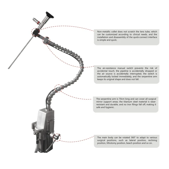
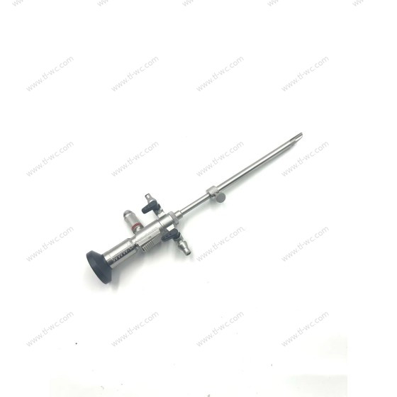
The shape and size fit the UBE surgical channel
UBE muscle stripping devices are usually designed to be relatively slender, with a length that can meet the requirements of puncturing the muscle tissue around the spine through the UBE surgical channel. This slender structure helps to reduce the space occupied in the surgical passage and avoid unnecessary compression on the surrounding tissues. Meanwhile, its width and thickness have also been meticulously designed to ensure that the muscle stripping operation has sufficient strength while not being too large in size to pass through the channel smoothly.
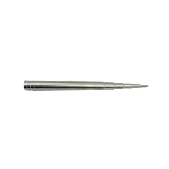
The diversity of head designs
Blunt head design: The head end of some muscle stripping devices is blunt, which can prevent accidental puncture of blood vessels or nerve damage during muscle stripping. Around the spine, the muscle tissue is closely adjacent to the nerves and blood vessels, and the blunt end can be safely separated along the direction of the muscle fibers. For instance, during cervical spine surgery, when stripping muscles such as the longus cervix, the blunt head end can effectively separate the muscles from the surface of the vertebral body without damaging the vertebral artery and cervical nerve.
Micro-arc or oblique head design: Some muscle stripping devices have a certain arc or Angle on their heads, which can better conform to the physiological curve of the spine and the anatomical form of the muscles. During lumbar spine surgery, the spine has physiological lordosis. The arc-shaped head can follow the curve of the lumbar spine, penetrate into the gap between the muscles and the vertebral body, and perform muscle dissection more precisely. Moreover, the bevel design enables the stripping device to function in different directions. For instance, when dealing with the lateral muscles of the spine, an appropriate Angle can make the operation more convenient.
Material characteristics
It is generally made of high-quality medical stainless steel or other materials with good biocompatibility and mechanical properties. This material has sufficient hardness to withstand the force during muscle separation, and at the same time, it has a certain degree of toughness to prevent fracture during the surgical process. Moreover, the material surface is smooth, facilitating sliding between tissues and reducing friction and damage to the tissues. UBE
The application scenarios of surgery
The application of cervical spine surgery
Anterior cervical surgery: In anterior cervical discectomy and fusion, a muscle discator is used to separate the muscles at the front of the vertebral body (such as the longus cervix, etc.) from the surface of the vertebral body. The muscle disintegrator is inserted through the UBE surgical channel. By taking advantage of the characteristics of its head end, the muscle is gently pushed away along the starting and ending points of the muscle and the direction of the fibers, providing a clear surgical field and operating space for subsequent surgeries (such as discectomy, intervertebral fusion device implantation, etc.). For instance, in anterior surgeries of the C4-C5 or C5-C6 cervical vertebrae segments, muscle strippers can effectively separate the muscles, reducing intraoperative bleeding and damage to the surrounding tissues.
Posterior cervical spine surgery: In posterior cervical spine surgery, such as spinal canal decompression or posterior fusion, a muscle disintegrator can be used to disintegrate the paraspinal muscles. Because the muscles at the back of the cervical vertebrae are relatively thin and there are many nerves, using an appropriate muscle stripping device can prevent nerve damage. First, insert the disintegrating device into the gap between the muscle and the lamina. Then, through operations such as rotation and sliding, separate the muscle from the lamina and the facet articular surface, creating conditions for the placement of spinal canal decompression or fusion surgical instruments.
Application in Lumbar spine surgery
Lumbar intervertebral disc surgery: In UBE surgery for lumbar intervertebral disc protrusion, a muscle disintegrator is used to expose the muscles around the intervertebral space. By stripping the erector spinae and other muscles in the waist, the intervertebral space can be better positioned and accessed. The design of its head end can help doctors quickly and effectively separate muscles without damaging nerve roots and blood vessels, providing convenience for surgeries such as nucleus pulposus removal. For instance, during L4-L5 or L5-S1 lumbar segment surgeries, a muscle stripping device can be used to strip the muscles around the intervertebral discs along the lateral or posterior side of the lumbar spine to expand the surgical field of view.
Lumbar decompression and fusion surgery: In the decompression surgery and lumbar fusion surgery for lumbar spinal stenosis, the muscle disintegrator is an indispensable instrument. It can separate the muscles around the spinal canal (such as multifidus muscles, etc.) from structures like the lamina, facet joints, and transverse processes. During decompression surgery, this helps to better expose the spinal canal and intervertebral foramen, facilitating the removal of hyperplastic bone mass and thickened ligamentum flavum. In fusion surgery, provide sufficient space for implanting the fusion device and performing bone grafting surgery.
Application in Thoracic surgery
Thoracic intervertebral disc surgery: In thoracic intervertebral disc protrusion surgery, a muscle stripping device is used to strip the muscle tissue between the thoracic vertebrae. Because the thoracic vertebrae are protected by the ribs and thorax, the surgical space is relatively narrow. The slender shape and appropriate head design of the muscle stripping device help it precisely strip muscles within a limited space. By separating the muscles from the vertebral bodies and intervertebral discs, it creates favorable conditions for the treatment of thoracic intervertebral discs.
Thoracic vertebra fracture surgery: During thoracic vertebra fracture surgery, a muscle stripping device can be used to separate the soft tissue around the fracture, facilitating the observation of the fracture and the operation of reduction and fixation. Separating the muscle from the fractured vertebral body can reduce the interference of the muscle on fracture reduction and avoid intraoperative damage to the nerves and blood vessels within the muscle.
Operation precautions and skills
Precautions for Operation
The importance of visualized operation: UBE surgery is performed under endoscopic visualization. Before using the muscle stripping device, please ensure that the endoscopic field of view is clear. Only by clearly understanding the relationship between the stripping device and the surrounding muscles, nerves, blood vessels and other tissues can a safe and effective surgery be carried out. Any blind operation may lead to complications such as tissue damage and bleeding.
Avoid excessive stripping and injury: When stripping muscles, avoid applying excessive force to prevent muscle tearing or damage to their blood supply. At the same time, it is necessary to pay attention to protecting nerves and blood vessels, especially in areas where muscles, nerves and blood vessels are closely intertwined, such as near the intervertebral foramen of the spine and beside the spinal canal. If you encounter a situation with significant resistance, do not forcibly peel it off. Instead, check the cause. It might be encountering tough fascia, vascular branches or nerve branches.
Cleaning and maintenance of instruments: Before the operation, it is necessary to check whether the head end of the muscle stripping device is intact, and whether there is any deformation or damage. After the operation, the instruments must be properly cleaned and disinfected to prevent residual tissues or bacteria inside the instruments from causing surgical infection and damage to the instruments. Operation
Technique
Insertion technique: Based on the anatomical path observed by the endoscope and the muscle distribution at the surgical site, slowly insert the muscle stripping device at an appropriate Angle and direction. During the insertion process, its slender shape can be utilized to carefully enter the gap between the muscle and the bone along the natural anatomical gap or the established working channel. For instance, during posterior lumbar spine surgery, when inserting a muscle stripping device, it can first be inserted along the gap beside the spinous process, and then the head end should be turned towards the gap between the lamina and the muscle.
Stripping technique: When performing muscle stripping, the movements should be gentle and gradual. Make the head end of the stripping device fully contact the edge of the muscle. Then, based on the starting point, ending point and fiber direction of the muscle, gradually separate the muscle from the bone surface through operations such as rotation and sliding. For more resilient muscles or areas with tight adhesion, the blunt end of the stripper can be used for initial separation first, and then, as needed, a head with a curvature or Angle can be used for more precise operations. Throughout the entire process, the position and operation method of the stripping device should be constantly adjusted in combination with the real-time observation of the endoscope to achieve the best stripping effect.
| Surgical instruments | |
| Material | Stainless steel |
| Cryogenic plasma | Autoclaving |

{{ item.content }}

{{ item.created_at_format }}
{{ item.reply }}
To find out more about our products and solutions, please fill out the form below and one of our experts will get back to you shortly.