Comprehensive analysis by single-portal endoscopy of the spine
I. Clinical Effects
As a core instrument in minimally invasive spinal surgery, the single-portal endoscope, with its feature of "single-channel integrated observation and operation", focuses on the demand for precise and minimally invasive treatment in clinical practice. Its core value is reflected in the following three aspects:
- Ultimate minimally invasive treatment, reducing tissue damage: The surgical operation can be completed through a single channel with a diameter of 5-8mm, without the need for additional incisions. It can minimize the stripping and traction of soft tissues such as muscles and fascia around the spine to the greatest extent. The intraoperative blood loss can be controlled within 5-10ml. The postoperative back pain score (VAS) of patients is generally lower than 3 points. Significantly reduce the risk of tissue trauma in traditional open surgery.
- Precisely adapted to the treatment of localized lesions: For localized lesions such as single-segment intervertebral disc protrusion (especially lateral recess type and intervertebral foramen type) and local spinal stenosis (such as thickening of the ligamentum flavum), single-portal endoscopy can directly reach the lesion area through the intervertebral foramen or laminar space approach, avoiding the interference of multi-channel operations on peripheral nerves and blood vessels. The incidence of surgical complications (such as nerve injury and rupture of the dural sac) is less than 1%.
- Shorten the postoperative recovery periodDue to the minimal trauma, patients can get out of bed and move around 6 to 8 hours after the operation, be discharged from the hospital 1 to 2 days after the operation, and resume normal work and life within 3 months after the operation. Compared with dual-channel surgery or open surgery, the recovery period is shortened by more than 50%, making it particularly suitable for elderly patients, those with underlying diseases, and people with high demands for recovery speed.
Ii. Design Considerations
The core design of a single-portal endoscope is to "achieve 'observation-operation' coordination within a single channel", which requires balancing the utilization rate of channel space, operational flexibility and organizational security. The specific design points are as follows:
- Material selectionThe lens body is made of medical-grade 316L stainless steel (outer layer) and high-transparency optical glass (lens part). 316L stainless steel is both corrosion-resistant (able to withstand sterilization at 134℃ high temperature and high pressure) and biocompatible, preventing the precipitation of metal ions. The optical glass adopts sapphire coating technology, with a light transmittance of over 98%, ensuring clear and distortion-free images.
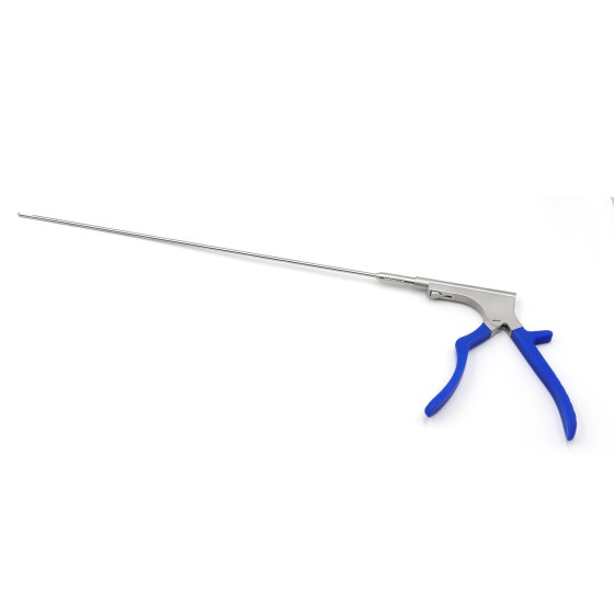
- Single-channel integrated layoutThe inner diameter of the channel is designed to be 5-8mm, and it is internally divided into an "optical zone" and an "operation zone". The optical zone is equipped with a high-definition CCD image sensor and an LED light source, achieving a 120° wide-angle field of view (covering the observation range required for spinal surgery). The operation area reserves 2 to 3 channels for micro-instruments, which can simultaneously accommodate grippers, radiofrequency electrodes, nucleus pulposus forceps and other instruments, and the operation of the instruments does not interfere with each other, meeting the requirement of "observation and operation at the same time".
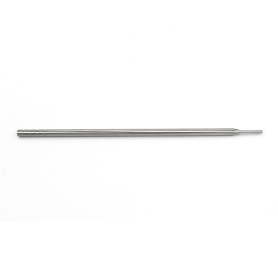
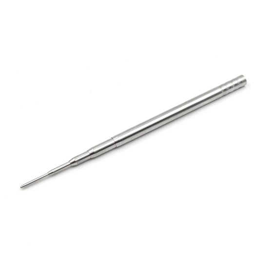
- Optimization of the mirror body shapeThe front end of the endoscope adopts a "diagonal cut" design (with an Angle of 15°-30°), which can conform to the anatomical curves of the intervertebral Spaces or foramina of the spine, avoiding the scraping of the bone at the edge of the vertebral body. The middle section of the mirror body transitions in a "tapering" style, gradually reducing the diameter from 12mm at the handle end to 8mm at the front end, thereby reducing the compression on the tissue during insertion.
- Positioning and control assistanceThe surface of the mirror body is engraved with millimeter-level scales (from 0mm at the front end to 50mm at the handle end), which helps doctors accurately determine the insertion depth. The handle is equipped with a "dual-knob control system", which respectively controls the Angle adjustment of the mirror body (±30° up and down, ±20° left and right) and the switch of the instrument channel. The operating torque is less than 0.5N · m, facilitating precise control by doctors with one hand.
- Ergonomic designThe handle adopts an "arc-shaped fitting" structure, which conforms to the natural grip arc of the palm. The surface is covered with anti-slip silicone material (friction coefficient ≥0.8) to prevent the instrument from sliding due to hand sweat during the operation. The weight of the handle is controlled at 150-200g, which makes it less likely for doctors to experience arm fatigue after long-term operation (1-2 hours) and enhances the stability of the operation.
Iii. Production Standards
As a Class II medical device, the production process of single-portal endoscopes must strictly follow the "Good Manufacturing Practice for Medical Devices" and international standards. The core standards include:
- Raw material control316L stainless steel shall comply with ASTM F138 standard (requirements for medical implantable grade stainless steel), and provide a material composition analysis report (such as chromium content 16-18%, nickel content 10-14%); Optical glass must pass the ISO 10993-1 biocompatibility test (cytotoxicity, allergenicity, irritability) to ensure no adverse reactions when in contact with human tissues.
- Production process specificationThe "precision CNC machining + laser welding" process is adopted, and the surface roughness (Ra) of the inner wall of the mirror body channel is ≤0.2μm, avoiding jamming when instruments enter and exit. The optical components adopt the "vacuum coating + precise assembly" process to ensure that the coaxiality error between the lens and the CCD sensor is ≤0.01mm, and there is no deviation in image transmission. Each device needs to undergo 200 simulated surgical operation tests to ensure smooth and error-free Angle adjustment and instrument channel switches.
- Sterility and packaging standardsIt adopts "high-temperature and high-pressure sterilization (134℃, 0.22MPa, 18 minutes) + double-layer aseptic packaging". The inner layer is medical dialysis paper (breathable and antibacterial), and the outer layer is aluminum-plastic composite film (puncture-resistant and moisture-proof). After sterilization, it needs to pass the EN ISO 11137 sterility verification to ensure that the sterility validity period is up to 2 years, and the packaging damage rate should be controlled within 0.1%.
- Quality system certificationThe manufacturing enterprise must pass the ISO 13485 medical device quality management system certification and the production license certification of the National Medical Products Administration (NMPA). Each piece of equipment must be accompanied by a "Product Qualification Certificate", "Sterilization Verification Report" and "User Manual", and the production batch, raw material source and quality inspection records can be traced through the unique product serial number.
Iv. Role in Spinal Endoscopic Surgery
Single-portal endoscopy is the "core carrier for observation and operation" in spinal surgery, running through the entire surgical process. Its specific functions are as follows:
- Single-channel establishment and organizational protectionAt the initial stage of the surgery, the doctor guides the single-portal endoscope into the lesion area through the intervertebral foramina or lamina space with a guide needle. The endoscope channel can expand the surrounding soft tissue, forming a stable "minimally invasive operation space", without the need for expander assistance, avoiding repeated stimulation of the tissue through multiple channels. Meanwhile, the rounded design at the front end of the endoscope can reduce the compression on the nerve roots and lower the risk of nerve damage.
- Real-time high-definition field of view transmission: The high-definition CCD sensor (resolution ≥1080P) and LED cold light source (adjustable brightness, 300-800lm) built into the endoscope can transmit real-time images of the surgical area (such as intervertebral discs, nerve roots, and dural sacs) to the display screen, with a magnification of up to 10-20 times. Help doctors clearly identify fine structures (such as nerve bundles and vascular branches) to avoid misoperation.
- Support for multi-device collaborative operation: The built-in operation channel of the endoscope can simultaneously accommodate 2 to 3 types of miniature instruments (such as 2.5mm diameter nucleus pulposus forceps and radiofrequency electrodes). Doctors can control them through the handle to complete operations such as "resection of diseased tissues (such as protruding nucleus pulposus), hemostasis (radiofrequency ablation), and annulus fibrosus repair" within the observation field without the need to replace instruments or adjust the position of the channel. Improve the efficiency of surgeries.
V. Working Principle
The working principle of a single-portal endoscope is based on a collaborative mechanism of "optical imaging + mechanical control + single-channel integration", as follows:
- Principle of Optical ImagingThe light emitted by the LED cold light source is conducted to the front end through the optical fiber inside the mirror body, illuminating the surgical area. The reflected light from the lesion area is focused by an optical lens (composed of 4 to 6 concave-convex lenses) and then transmitted to the CCD image sensor. The sensor converts light signals into electrical signals, which are then transmitted to the image processing system via data lines. Eventually, high-definition color images are presented on the display screen, enabling "real-time observation".
- Single-channel operation principleThe operation channels inside the mirror body adopt a "concentric circle" design, with the optical area at the center (occupying 1/3 of the channel diameter) and three evenly distributed instrument channels on the periphery (each occupying 1/3). When the instrument is inserted, it can slide along the inner wall of the channel. The "guide groove" design of the channel (with a depth of 0.1mm) can restrict the movement direction of the instrument, ensuring that the instrument is always within the observation field of view and achieving "synchronization of observation and operation".
- Principle of Angle adjustmentThe Angle adjustment knob at the handle is connected to the front end of the mirror body through a "micro gear transmission mechanism". When the knob is turned, the gear drives the "universal joint" at the front end of the mirror body to rotate, achieving the adjustment of the up and down and left and right angles. The transmission mechanism adopts "precision ball bearings" to ensure no jamming during adjustment, with an Angle error of ≤1°, helping doctors precisely align with the lesion area.
Vi. Usage Steps
The use of single-portal endoscopes must strictly follow the process of "precise positioning - minimally invasive operation - postoperative care", and the specific steps are as follows:
- Preoperative preparation
- Instrument inspectionThe nurse needs to inspect the appearance of the endoscope (the endoscope body has no deformation or cracks, and the lens has no stains), connect the display screen to test the image (the clarity and color have no deviation), and check the instrument channel (insert the grips and radiofrequency electrodes to ensure smooth sliding). Confirm that the aseptic packaging is undamaged and the sterilization validity period is within the specified range.
- Patient preparationThe patient underwent preoperative CT or MRI examination to determine the lesion segment (such as L4-L5 intervertebral disc protrusion) and location (lateral crypt type). On the day of the operation, the patient was placed in a prone position. Soft pillows were placed on the chest and pelvis (to keep the spine in a neutral position). The surgical area (back) was disinfected with iodophor (range: 3 vertebrae above and below the lesion segment, left and right to the midaxillary line). A sterile surgical towel was spread and local anesthesia (0.5% lidocaine) was used.
- Intraoperative operation
- Guide pin positioningUnder the X-ray fluoroscopy of the C-arm X-ray machine, a 2.0mm diameter guide needle is punctured into the lesion area through the intervertebral foramen approach (or the interlaminal space approach), and the position of the guide needle is confirmed to be accurate (≤2mm from the protruding nucleus pulposus).
- Endoscope insertionSlowly insert the single-portal endoscope (with an outer sleeve diameter of 6mm) along the direction of the guide pins. Confirm the insertion depth (usually 8-12cm) through the scale on the surface of the endoscope body. Under fluoroscopy, ensure that the front end of the endoscope body is aligned with the lesion area and fix the position of the endoscope.
- Lesion managementTurn on the endoscope light source and image system. Under the field of view of the display screen, insert the nucleus pulposus forceps through the operation channel and gradually remove the protruding nucleus pulposus tissue. If bleeding occurs during the operation, insert a radiofrequency electrode for hemostasis (with the temperature controlled at 60-80℃). During the operation, adjust the Angle of the lens body through the handle to ensure clear observation of the nerve roots and dural sac throughout the process and avoid damage.
- Surgical conclusionConfirm that the lesion tissue has been thoroughly removed (no residue observed under fluoroscopy and endoscopy), remove all instruments, slowly withdraw the endoscope, and apply sterile dressings at the puncture site (no suturing required).
- Postoperative management
- Cleaning and disinfection of instruments: After use, the endoscope should first be rinsed with running water (to remove blood and tissue residues), then soaked in an enzyme cleaner for 30 minutes (concentration 1:100), ultrasonically cleaned for 15 minutes (frequency 40kHz), and finally subjected to high-temperature and high-pressure sterilization (in accordance with production standards). After sterilization, it should be stored in a dedicated instrument cabinet (temperature 20-25℃). Humidity: 40-60%.
- Patient observation and rehabilitationAfter the operation, monitor the patient's vital signs (blood pressure, heart rate) and the sensory and motor functions of the lower extremities (such as dorsiflexion and plantar flexion muscle strength), and observe whether there is bleeding or redness and swelling at the puncture site. Guide the patient to get out of bed and move around 6 hours after the operation (wearing a waist support), avoid bending over and sitting for long periods within one week after the operation, and perform functional exercises for the lumbar and back muscles (such as the five-point support method) within one month after the operation.
Vii. Precautions
To ensure the safety and effectiveness of single-portal endoscopes, the following precautions must be strictly followed:
- Preoperative precautions
- Assessment of contraindicationsClarify surgical contraindications, including: Patients with multi-segment intervertebral disc protrusion (such as simultaneous protrusion of L3-L4 and L4-L5), severe spinal instability (such as grade II or above vertebral spondylolisthesis), infection in the puncture path (such as local skin abscess), and coagulation dysfunction (such as platelet < 100×10⁹/L) are not suitable for single-portal endoscopic surgery to avoid intraoperative risks.
- Equipment compatibility checkConfirm that the interfaces of the endoscope are compatible with those of the display screen, RF instrument and other devices (such as HDMI interface, high-frequency connector), and test the interactivity of the equipment (such as no interference to the endoscope image when the RF electrode is working) to avoid surgical interruption due to equipment incompatibility.
- Intraoperative precautions
- Puncture and insertion controlGuide needle puncture and endoscope insertion should be carried out under the full guidance of fluoroscopy. The position should be confirmed once every 0.5cm of advancement to avoid excessive puncture leading to rupture of the dural sac. When inserting the endoscope, do it gently. If you encounter resistance (such as bone obstruction), adjust the Angle before pushing. Violent insertion is strictly prohibited.
- Vision and operation managementThroughout the operation, maintain a clear field of vision. If the field of vision is blurred (such as blood obstruction), normal saline (pressure ≤30mmHg) should be injected through the operation channel for rinsing, or an aspirator should be used for cleaning. When operating the instrument, ensure that the front end of the instrument is always within the field of vision, avoid blind probing (especially near the nerve roots), and control the operation force within 5-10N to prevent the instrument from breaking.
- Aseptic operation is strictly carried outAfter the endoscope is inserted, its sterile part (from the front end of the endoscope body to 10cm of the handle) must not come into contact with non-sterile items (such as the edge of the operating table, the outside of the doctor's gloves). When changing instruments, the entrance of the instrument passage should be wiped with sterile gauze to prevent contaminants from entering the passage. Surgical personnel need to change sterile gloves regularly to ensure that the sterile area is not contaminated.
- Postoperative precautions
- Equipment maintenance and upkeepAfter using the endoscope, avoid collision (especially the lens part). When cleaning, do not use a hard-bristled brush to wipe the lens (special lens paper should be used instead). Regularly (every 50 uses) conduct performance tests (image clarity, Angle adjustment accuracy). If lens scratches or channel jamming are found, send it to the manufacturer for repair in time. Do not continue to use it.
- Monitoring of patient complicationsWithin 24 hours after the operation, pay close attention to whether there is "postoperative reactive pain" (caused by nerve root edema, mannitol dehydration can be given) and puncture site infection (such as redness, swelling, heat and pain, antibiotic treatment is required). Be vigilant for delayed dural sac leakage within one week after the operation (such as headache and dizziness, bed rest and fluid replacement are required). Any abnormalities should be dealt with promptly.
- Contraindications of rehabilitation guidanceInform the patient to avoid strenuous exercise (such as running and weightlifting), bending over to lift heavy objects (weight ≤5kg), and prolonged bending over for work (such as mopping and sweeping) within three months after the operation. If numbness in the lower limbs worsens or incontinence occurs, medical attention should be sought immediately to rule out the risk of nerve damage.