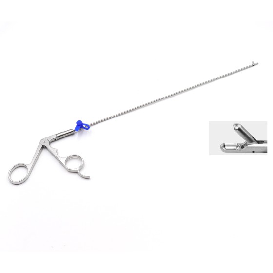
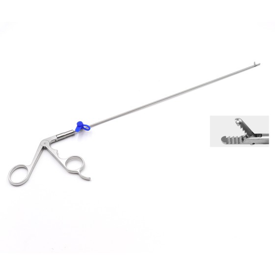
Menu

Spinal endoscopic surgical instrument eccentric guide rod expansion tube

Quantity:
Sales: 236
SKU:{{ product.sku }}
Model: {{ product.model }}
weight: {{ product.weight }} Kilogram

{{ variable.name }}

{{ value.name }}

The following is an introduction to the eccentric guide rods and dilation tubes in spinal endoscopic surgical instruments

Eccentric guide rod

    • Structural design
      • ShapeIt is usually a cylindrical structure with a cone. The cone is set at the end of the cylinder, and the end is a blunt cone structure, which can ensure that the guide rod will not damage important structures such as nerves, dural sacs, and blood vessels. Its cross-section can be circular or elliptical. The length of the circular eccentric guide rod ranges from 230mm to 400mm, the outer diameter from 5mm to 10mm, the diameter of the inscribed circle from 2mm to 6mm, and the outer diameter of the blunt conical tip from 2mm to 3mm. The length of the elliptical eccentric guide rod is between 140mm and 400mm, the long axis is between 6mm and 10mm, the short axis is between 4mm and 8mm, the inscribed circle is located at one end of the long axis, the diameter of the inscribed circle is between 2mm and 6mm, and the outer diameter of the elliptical conical tip is between 2mm and 3mm.
      • HandleThe eccentric guide rod is equipped with a handle, which is sleeved on the eccentric guide rod. There are two through holes of different diameters on it, corresponding to the structure of the eccentric guide rod. It can be applied to eccentric guide rods of different diameters and is convenient for manual rotation of the eccentric guide rod. At the end where the handle is provided, a planar structure is also provided for the convenience of fixing the handle.
      • Eccentric grooveThe eccentric guide rod is provided with an eccentric groove for placing the guide rod. The guide rod and the eccentric guide rod are in a semi-embedded structure, which facilitates the installation and rotational fit of the two guide rods.
    • Working principleIn spinal endoscopic surgery, the approximate position of the surgery is first determined by placing a guide rod, and then the eccentric guide rod is fitted to the outside of the guide rod. Since the central axes of the eccentric guide rod and the guiding guide rod are parallel, and as the eccentric guide rod rotates, the distance between the two central axes gradually changes, by rotating the guiding guide rod or the eccentric guide rod, the axial distance between the two guide rods can be changed, thereby adjusting the position of the surgical instruments and enabling them to reach the surgical site more accurately2
    • Clinical application advantages
      • Improve positioning accuracyIt can more precisely guide surgical instruments to the lesion site, reducing surgical errors. For some deep and difficult-to-reach lesion tissues, the eccentric adjustment function of the eccentric guide rod can effectively solve the positioning problem2
      • Reduce the difficulty of the surgeryDoctors no longer need to repeatedly adjust the position of the guide rod under X-ray fluoroscopy as in traditional methods. One or two adjustments can reach the correct surgical site, greatly reducing the difficulty of spinal endoscopy surgery and relatively lowering the technical requirements for the surgeon2
      • Reduce radiation exposureIt reduces the number of times doctors and patients are exposed to X-rays, and lowers the damage of radiation to the body2
      • Increase operational flexibilityIn intervertebral foramen endoscopic surgery, the cannula can be rotated in different directions to grind bone in different directions, and multiple grinding operations can be performed in the same direction to better control the grinding range, ensuring the accurate placement of the working cannula and the wide viewing Angle of the endoscope. This provides a larger operating space for the working cannula within the spinal canal, allowing it to be inclined towards the head end, tail end, and dorsal side3

Expansion tube

    • Structural features
      • Diverse sizesThere are different diameter and length specifications. The common diameter range is from a few millimeters to tens of millimeters, and the length is generally around 130mm to 160mm to adapt to different surgical sites and surgical requirements. For example, there is an expansion tube (10 to 7) 10.0160 inner hole 7.0, expansion tube 14.3There are various specifications such as 160 inner hole 7.0, expansion tube 14.3*130 inner hole 10.0, etc1
      • Shape characteristicsIt is generally cylindrical in shape, and the front end may be conical, which is convenient for insertion into tissues and step-by-step expansion of soft tissue channels. The edge of the partial expansion tube has a through eccentric inscribed circle, with the diameter of the inscribed circle being ≥4.1mm5
      • Material propertiesIt is usually made of medical-grade metals or polymer materials, featuring excellent strength, toughness and biocompatibility. It can maintain a stable shape during the process of tissue expansion without causing adverse effects on human tissues.
    • Mechanism of actionIn spinal endoscopic surgery, the expansion tube needs to be gradually inserted along the guide wire or guide rod. Through the step-by-step expansion, the skin, muscles, fascia and other soft tissues are stretched out to form a channel, so that subsequent surgical instruments such as working cannulas and endoscopes can smoothly enter the surgical site, reducing damage to the surrounding tissues and providing sufficient space for the surgical operation3
    • Clinical application significance
      • Establish a surgical passageIt is a key instrument for establishing a surgical channel, providing a necessary path for subsequent surgical operations, enabling endoscopes and other surgical instruments to smoothly reach the lesion site for observation and treatment.
      • Protect the surrounding organizationsThe step-by-step expansion approach enables soft tissues to gradually adapt to the expansion process, reducing tearing and damage to surrounding tissues, lowering the risk of surgical complications, and facilitating postoperative tissue recovery.
Upload a JPG or PNG image. 300 x 300 is recommended.
Only jpg/jpeg/png/mp4/mov files can be uploaded, and it should not exceed 50M
Submit Cancel
Add Reviews
Add Reviews
{{ item.user_name_format }}

{{ item.content }}

{{ item.created_at_format }}

Reply:

{{ item.reply }}

Related Products
Spinal endoscope - pulposus forceps - 3.5mm x 330mm -0° long cup
SELF
Spinal endoscope - pulposus forceps - 3.5mm x 330mm -0° long cup
please To check the price
Sales:686
Spinal endoscope - nucleus pulposus forceps with toothed gripper - 2.5mm x 330mm -0°
SELF
Spinal endoscope - nucleus pulposus forceps with toothed gripper - 2.5mm x 330mm -0°
please To check the price
Sales:782
Spinal endoscope - nucleus pulposus forceps - 3.0mm x 330mm -0° long cup
SELF
Spinal endoscope - nucleus pulposus forceps - 3.0mm x 330mm -0° long cup
please To check the price
Sales:555