Spinal Endoscope & Minimally Invasive Surgical Instruments Manufacturer | QuNaMai
Wishlist

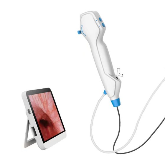
{{ variable.name }}


Structural features and advantages
Material and process advantages
High-quality stainless steel material: The UBE cervical biting pliers are made of high-quality stainless steel. This material has high strength and can withstand the force generated by the occlusal tissue during cervical spine surgery, and is not prone to deformation or damage. Its excellent corrosion resistance also ensures that the equipment can maintain good performance after long-term use and contact with various body fluids and disinfectants. A perfect production process system ensures that each bite force can meet high-quality standards, making its functions more reliable. For instance, in the complex anatomical environment of the cervical vertebrae, high-quality occlusal forceps can perform multiple occlusal operations stably without the forceps head loosening or breaking due to material or manufacturing issues.
Overload protection device: The overload protection device is a major highlight of this equipment. During cervical spine surgery, occlusal forceps may encounter various tissues of different hardness and toughness, such as tough cervical ligaments and osteophytes. Overload protection devices can effectively prevent the instruments from being damaged due to excessive force. This not only extends the service life of the instruments, reduces the cost of hospital equipment renewal, but also ensures the continuity of operations during the surgical process. When biting onto harder tissues, the overload protection device will automatically limit the biting force to prevent damage to the jaws, thus ensuring that each biting operation can be completed smoothly.
The design advantages that conform to the UBE surgical concept
Compact and exquisite size design: UBE surgery emphasizes the use of compact and exquisite instruments for precise operations. The size design of 2.5MM×180MM fully conforms to this concept. In cervical spine surgery, the anatomical structure of the cervical spine region is very complex, with dense distribution of peripheral nerves and blood vessels, and the surgical operation space is relatively narrow. The 2.5MM diameter enables the occlusal forceps to smoothly pass through the UBE surgical channel and enter the cervical vertebrae, reducing compression and damage to the surrounding tissues. The 180MM length provides sufficient extension range for the surgical operation, facilitating the doctor to perform occlusion at the appropriate operating position and better adapting to the surgical requirements of different cervical vertebrae segments and depths. For instance, in the surgery for cervical intervertebral disc protrusion, these small occlusal forceps can accurately reach the intervertebral space and occlude the lesion tissue around the protruding nucleus pulposus without interfering with the surrounding nerve roots and blood vessels.
The design features of the inverted tooth gripper: The design of the inverted tooth gripper has unique advantages in cervical spine surgery. Inverted teeth can enhance the grip force of the occlusal forceps on tissues. During the biting and cutting process, especially for some soft or easily sliding tissues, such as the nucleus pulposus tissue of the cervical intervertebral disc and fragments of degenerated annulus fibrosus, inverted teeth can effectively prevent tissues from sliding out of the forceps, which is conducive to accurately biting the target tissue and improving the efficiency of the surgery. For instance, when removing free nucleus pulposus fragments from the cervical spinal canal, the inverted tooth gripper can firmly grasp the fragments and then perform biting or removal operations to prevent the fragments from moving within the spinal canal and causing secondary damage to the nerves.
Application scenarios of cervical UBE surgery
Surgery for cervical intervertebral disc protrusion
In the UBE surgical treatment of cervical intervertebral disc protrusion, the biting forceps play an important role. Firstly, after the biting and cutting forceps locate the protruding nucleus pulposus tissue through endoscopy, they accurately reach the intervertebral space by taking advantage of their compact size and the inverted tooth grasping forceps design. The inverted teeth can grasp the protruding nucleus pulposus and the surrounding degenerated annulus fibrosus tissue that may compress the nerve. Then, the biting and cutting operation is performed to gradually remove these diseased tissues. During this process, the overload protection device can ensure that the instrument will not be damaged when biting and cutting harder annulus fibrosus tissue. The high-quality stainless steel material guarantees the sharpness and stability of the clamp head, making the biting and cutting process more precise and efficient, thereby reducing the compression on the nerve roots and alleviating the patient's upper limb pain, numbness and other nerve compression symptoms.
Cervical spinal canal decompression surgery
In UBE surgery for cervical spinal stenosis, occlusal forceps are used to bite off the tissue that causes spinal stenosis. For instance, in cases of hypertrophy of the ligamentum flavum within the spinal canal, the occlusal forceps can smoothly enter the spinal canal through their 2.5MM diameter. Then, the ligamentum flavum tissue is grasped using the inverted tooth gripper, followed by occlusion and incision to expand the volume of the spinal canal. For the parts of the cervical lamina accompanied by bone hyperplasia, occlusal forceps can also precisely bite off the hyperplastic bone without damaging the spinal cord and nerve roots. Throughout the entire spinal canal decompression process, the compact and exquisite design enables the occlusal forceps to operate flexibly within the complex anatomical structure of the cervical vertebrae, while the perfect production process and overload protection device ensure the safety and effectiveness of the surgery, providing a reliable guarantee for the patient's nerve decompression.
Cervical fusion surgery assistance
During the preparation stage of cervical spine fusion surgery, occlusal forceps can be used to clean the soft tissues and part of the bone at the fusion site. For instance, when dealing with the small joints of the cervical vertebrae, the occlusal forceps can use the reverse tooth gripper to grasp the cartilage tissue and a small amount of bone on the surface of the small joints, and then perform occlusion and incision to create a favorable bone surface environment for the implantation of the fusion device. Its compact size ensures that the surrounding nerves and blood vessels will not be damaged during operation. Meanwhile, the high-quality stainless steel material and overload protection device can ensure that the occlusal forceps can work stably when handling tissues of different hardness, increase the success rate of the surgery, and promote the smooth progress of cervical fusion surgery.
Operation precautions and skills
Precautions for Operation
Since UBE surgery is performed under endoscopic visualization, it is essential to ensure a clear endoscopic field of view before the operation to accurately observe the relationship between the occlusal forceps and the surrounding tissues. During the insertion and operation of the occlusal pliers, special attention should be paid to avoiding damage to important tissues such as nerves and blood vessels. The nerves and blood vessels in the cervical region are extremely dense. A slight mistake may lead to serious consequences. At the same time, it is necessary to check whether the overload protection device is working properly to avoid damage to the equipment or injury to the patient due to device failure.
Operation skills
Insertion technique: Based on the anatomical path observed by the endoscope and the structural characteristics of the cervical vertebrae at the surgical site, insert the occlusal forceps at an appropriate Angle and direction. By taking advantage of its compact size, it can be slowly inserted along natural anatomical gaps or established working channels, avoiding forced insertion that may damage the surrounding tissues.
Occlusal technique: When occluding tissues, first gently bring the jaws of the occlusal pliers close to the target tissue. Use the inverted tooth gripper design to allow the tissue to fully enter the jaws. Then, adjust the occlusal force appropriately according to the size, texture, and toughness of the tissue. For softer tissues, a smaller bite force can be applied to prevent excessive compression and slippage out of the jaws. For harder tissues, such as bone hyperplasia, the biting force can be gradually increased within the range allowed by the overload protection device. Throughout the entire surgical procedure, the position and operation method of the cutting forceps should be flexibly adjusted in combination with the real-time observation of the endoscope to achieve the best surgical outcome.
| Surgical instruments | |
| Material | Stainless steel |

{{ item.content }}

{{ item.created_at_format }}
{{ item.reply }}
-560x560.png)
-560x560.png)
-560x560.png)
To find out more about our products and solutions, please fill out the form below and one of our experts will get back to you shortly.