
Percutaneous Endoscopic Lumbar Discectomy
Author: QuNaMai
Release time: 2025-09-20 05:39:48
View number: 29485
Case of Transforaminal Endoscopic Treatment for High-Level Massive Lumbar Disc Herniation
I. Case Overview and Surgery Summary
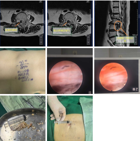
The patient was transferred from another hospital and diagnosed with high-level massive lumbar disc herniation. Their pain worsened after conservative treatment—they even "could not sleep at night due to severe pain, often clutching their thigh". After evaluation, we decided to perform minimally invasive transforaminal endoscopic surgery. Before the operation, we clearly told the patient that "a small hole surgery can relieve the pain", which eased their anxiety.
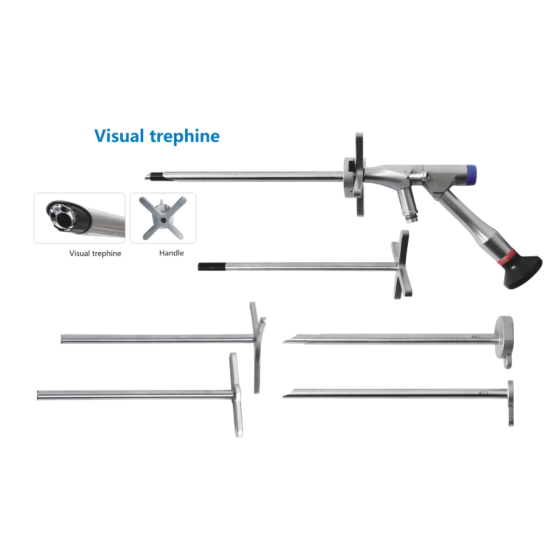
The surgery was conducted with a small posterolateral incision under C-arm fluoroscopy guidance. The core equipment used was the QuNaMai transforaminal endoscopic system. This system provides high-definition visualization of details inside the spinal canal, helping us clearly distinguish between the herniated nucleus pulposus and nerve roots to avoid accidental injury. The matching adjustable working channel can be flexibly adjusted according to the narrow space of the high-level intervertebral foramen, making the operation convenient. During the surgery, the patient reported immediate pain relief, and the entire process was completed with relaxed communication.
II. Key Surgical Techniques
1. Foraminal Preparation and Working Channel Establishment
First, a water-cooled burr (QuNaMai brand) was used to grind down the hypertrophic zygapophyseal joints to create sufficient space. This burr cuts quickly without generating excessive heat, preventing damage to surrounding bone tissue. Next, a disposable sterile working channel was inserted—it has a smooth inner wall and a blunt tip, so it does not touch the dural sac, ensuring high safety.
2. Herniated Tissue Removal and Repair
QuNaMai double-jointed nucleus pulposus forceps were used to remove the massive herniated tissue in pieces. This forceps can flexibly reach into the narrow high-level spinal canal; its jaw edges are polished smooth, avoiding tearing of the nerve roots. At the same time, a suction tube was used to timely remove blood and tissue debris, maintaining a clear surgical field. Finally, a dual-frequency radiofrequency electrode was used for hemostasis and to shrink and repair the torn annulus fibrosus. The electrode has a curved tip that fits closely to the surface of the annulus fibrosus, achieving better repair results.
3. Tips for Handling Massive Herniated Tissue
For such a large herniated mass, we did not attempt to remove it all at once. Instead, we used appropriately sized nucleus pulposus forceps to first remove the free part in the spinal canal, then clear the remaining tissue in the intervertebral disc. We first used a thin probe to gently separate adhesions between the herniated tissue and nerve roots, and finally used a dissector to release surrounding adhesions, ensuring complete decompression of the nerve roots.
III. Key Points of Team Collaboration
In the surgical team, nurses carefully checked the entire set of QuNaMai instruments (including endoscopes, forceps, and consumables) before the operation. During the surgery, they prepared tools in advance according to the procedure—for example, adjusting the burr before use and checking the stability of the radiofrequency electrode connection—to ensure a smooth surgical rhythm.
IV. Experience and Future Suggestions
- Instrument selection is crucial for high-level spinal surgery. QuNaMai’s transforaminal endoscopes and forceps are compact, suitable for operations in narrow spaces. The disposable consumables also help prevent infection, making them reliable to use.
- In the future, we can refer to the instrument set concept of professional platforms, integrating small endoscopes, thin probes, and forceps of different sizes needed for high-level surgery into a complete set. This way, there is no need to count instruments one by one before surgery, saving a lot of time.
Related Products
Spinal endoscope - single portal endoscope - 6.3mm x 181mm -30° instrument channel 3.75MM
$6,500.00
$8,000.00
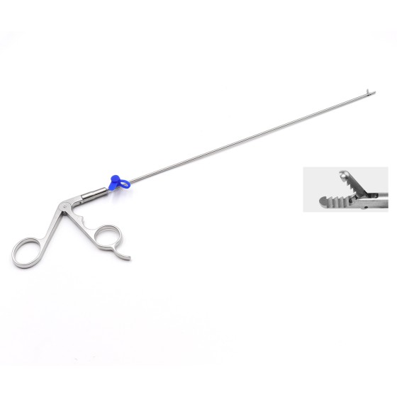
Spinal endoscope - nucleus pulposus forceps with toothed gripper - 3.5mm x 260mm -0°
$500.00
$600.00
Spinal endoscope - nucleus pulposus forceps with toothed gripper - 2.5mm x 330mm -0°
$500.00
$600.00
Spinal endoscope - nucleus pulposus forceps with toothed gripper - 3.5mm x 330mm -0°
$500.00
$600.00